
Normal fundus.

Fundus showing Macular edema.

Central Retinal Vein Occlusion (CRVO)
.
.
Series of fundus illustrations. The fundus is the interior of the eye. Some of the anatomy in the fundus includes the retina, the macula and optic disc. The first illustration shows healthy anatomy of the left eye, the next shows slight macular edema, and the bottom figure shows a condition called central retinal vein occlusion (CRVO). These disorders could lead to blurred vision or loss of vision.
Patient scans were used as primary references for this series.

Cross-section of a normal fovea.
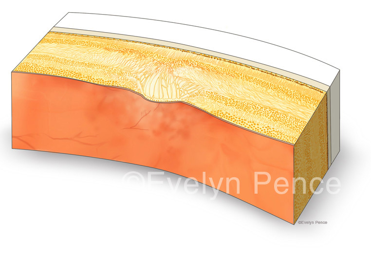
Cross-section with macular edema.
Block diagrams comparing a cross-section of a normal fovea to one with macular edema.